
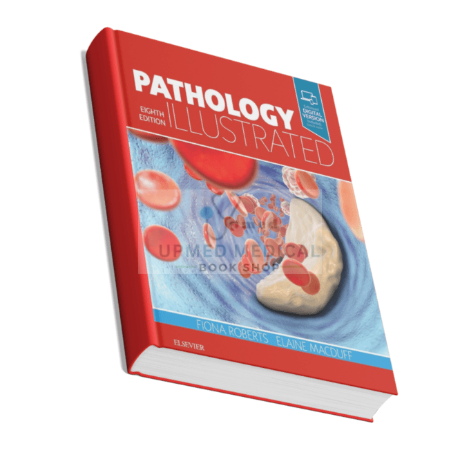
Pathology Illustrated
₨ 1,573 ₨ 3,155Price range: ₨ 1,573 through ₨ 3,155
- Author(s): Fiona Roberts, Elaine MacDuff
- Edition / Volume: Eighth Edition
- Publisher: Elsevier
Pathology Illustrated provides a vibrant visual guide to pathological processes with essential clinical correlations. This eighth edition is an indispensable resource for mastering the fundamentals of disease mechanisms and their practical applications in medicine.
About the Book
Pathology Illustrated stands as a definitive visual resource for understanding the complex mechanisms of disease. Designed for clarity and retention, this edition bridges the gap between foundational pathological processes and their real-world clinical correlations. Each illustration is carefully crafted to simplify intricate concepts, making it easier for learners to grasp the structural and functional changes that occur in disease states.
The book systematically covers a wide spectrum of pathological conditions, emphasizing the relationship between morphological changes and clinical presentation. By integrating visual learning with concise explanatory text, it supports both rapid review and deep comprehension. This approach is particularly valuable for students and practitioners who need to connect theoretical knowledge with diagnostic and therapeutic decision-making.
Key Themes & Topics Covered
- Fundamentals of cellular injury and adaptation
- Inflammation and repair mechanisms
- Neoplasia and tumor biology
- Systemic pathology with clinical correlations
- Diagnostic imaging and histopathological findings
- Integration of pathophysiology with clinical practice
Who Should Read This?
This volume is ideal for medical students, pathology residents, and healthcare professionals seeking a clear, image-driven reference. It also serves as a valuable review tool for those preparing for board examinations or clinical rotations where a strong grasp of disease mechanisms is essential.
Add Pathology Illustrated to your professional library and strengthen your visual mastery of disease processes.
| Weight | N/A |
|---|---|
| Print Type | Black & White, Color Matt Finish |
Be the first to review “Pathology Illustrated”
Related products





Reviews
There are no reviews yet.